When Is a CT Scan or Nasal Endoscopy Needed Before Revision Rhinoplasty?

When Is a CT Scan or Nasal Endoscopy Needed Before Revision Rhinoplasty?
Key answer
Before revision rhinoplasty, CT and nasal endoscopy are not required for every patient; they are supplementary tools used when the physical examination alone cannot answer a question. Nasal endoscopy evaluates the inner lining, the septum, and the nasal passages, while CT shows bony structure, the position of implants or cartilage grafts, and changes in surrounding tissue. They are considered particularly when there is nasal obstruction, suspected inflammation around an implant, or uncertainty about the materials and extent of the previous surgery. Whether any test is needed, and which one, varies by individual and is decided after an in-person examination.
What does the examination check before any test is ordered?
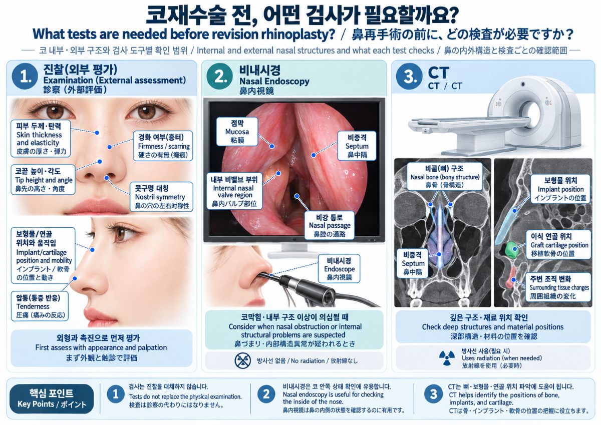
Testing does not replace the examination; it answers questions the examination raises. In revision cases the examination generally covers:
- Tip height and angle, and symmetry of the nostrils
- Skin thickness, elasticity, and any firmness or hardening
- On palpation, the position, mobility, and tenderness of an implant or graft
- Breathing, including where and how the obstruction occurs (such as at the nasal valve area)
- Records of previous surgery (materials, donor sites, number of operations) and how symptoms have changed over time
If breathing symptoms or internal abnormalities are suspected, endoscopy is considered; if deeper structures such as bone, an implant, or calcification need to be assessed, CT is considered. Conversely, when concerns are limited to appearance, the previous surgery is well documented, and the examination is sufficient, planning may proceed without additional tests.
What do nasal endoscopy and CT each show?
| Nasal endoscopy | CT | |
|---|---|---|
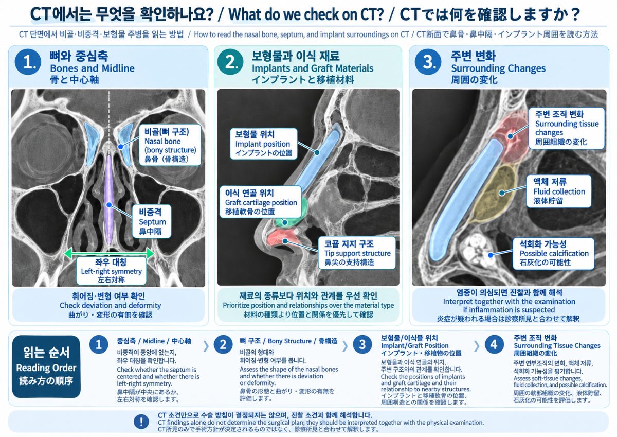
| What it evaluates | Inner mucosa, septum, nasal passages, nasal valve area | Bony structure (nasal bones, maxilla), septum, position of implants or grafts, changes in surrounding tissue |
| Typically considered when | Nasal obstruction, mucosal assessment, suspected internal deformity | Previous materials or structure unclear, suspected abnormality around an implant, bony deformity |
| Radiation | None | Yes (limited to what is necessary) |
| Limitations | Deep structures behind the mucosa are not directly visible | Fine mucosal detail and soft-tissue quality are limited |
On CT, clinicians can look for displacement of an implant, fluid collection or calcification around it, and the presence and location of cartilage grafts. Imaging findings must be interpreted together with the examination; images alone do not determine the treatment plan. In some cases, ultrasound is used as a supplementary way to assess the area around an implant.
Which findings lead to additional testing?
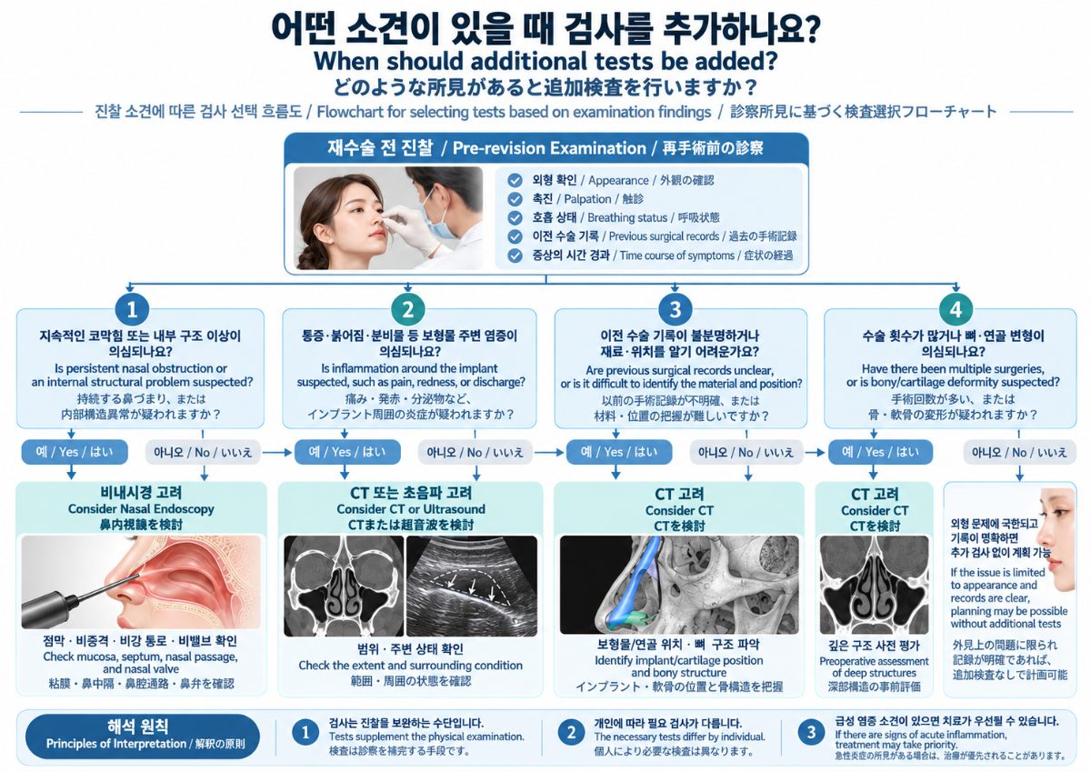
The following situations commonly prompt additional testing. They indicate a general direction only; whether they apply to an individual depends on the examination.
- Persistent nasal obstruction: endoscopy may be considered to look for internal causes such as septal deviation, mucosal thickening, or narrowing at the nasal valve area.
- Pain, redness, or discharge suggesting inflammation around an implant: CT or ultrasound may be considered to assess the extent and the area around the implant; when acute signs are present, treatment may take priority over testing. The distinction between inflammatory and scar-related contracture is discussed in How Do Inflammatory Contracture and Scar Contracture Differ?.
- Unclear surgical history: if it is not known what material is placed where, the surgical plan itself changes, and CT may help.
- Multiple prior operations, or suspected bone or cartilage deformity: CT may be considered to estimate the amount and condition of available cartilage and the bony framework.
- Appearance-only concerns with a clearly documented history: the examination may be sufficient, and no additional test is performed.
Test results can also influence which materials and techniques are chosen or avoided. Criteria for deciding whether to remove an implant are covered in When Does an Implant Need to Be Removed in Revision Rhinoplasty?, and how a simply upturned tip differs from a contracted nose is covered in How Do You Tell a Simply Upturned Nose from a Contracted Nose?.
How does the decision process work in practice?
As an anonymized, general line of reasoning: when a patient describes a tip that has gradually risen and skin that feels firmer, the examination first checks tip angle, skin hardening, and tenderness. If findings such as pain or discharge raise the possibility of inflammation, imaging of the area around the implant is considered; if breathing difficulty accompanies it, endoscopy evaluates the inside of the nose. If no records of the previous surgery exist, CT can help identify the materials and their positions before techniques and materials are chosen. If the concern is limited to appearance and the history is clear, surgery may be planned without further tests. This describes the order of reasoning, not the course of any particular patient.
For timing considerations, see also When Can Revision Rhinoplasty Be Performed After the First Surgery?.
Summary
- Pre-operative testing before revision rhinoplasty is a supplementary step used when the examination leaves open questions.
- Nasal endoscopy looks at the inner lining, septum, and passages; CT shows bone, the position of implants or cartilage, and surrounding changes.
- CT uses radiation, so it is used only when justified and within a limited range; it is not performed for every patient.
- Imaging is interpreted together with the examination; the necessary tests and treatment plan vary by individual and require in-person assessment.
Frequently asked questions
Q. Is a CT scan always required for revision rhinoplasty?
A. No. When the previous surgery is well documented and the examination is sufficient, planning may proceed without CT. It is considered when materials or structure are unclear, or when inflammation or bony deformity is suspected.
Q. Is nasal endoscopy painful?
A. A thin scope is used to view the inside of the nose; discomfort varies between individuals. A topical anesthetic may be used when needed.
Q. I am concerned about CT radiation.
A. CT uses radiation, so the principle is to perform it only when there is a clear need and within a limited range. Whether it is needed is best discussed after an examination.
Q. What if I have no records from my previous surgery?
A. Without records, the type of implant or whether cartilage was harvested may be unknown; imaging and examination can clarify what is possible, and some aspects may only be confirmed during surgery.
Q. If the tests show no abnormality, does that mean no revision is needed?
A. Normal results do not exclude surgery being considered for appearance or symptoms, and abnormal findings do not always require surgery. Testing is one element of the decision.
Related columns
- How Do Inflammatory Contracture and Scar Contracture Differ?
- When Does an Implant Need to Be Removed in Revision Rhinoplasty?
- How Do You Tell a Simply Upturned Nose from a Contracted Nose?
- When Can Revision Rhinoplasty Be Performed After the First Surgery?
Sources
- Neligan PC (ed.), *Plastic Surgery*, Elsevier — rhinoplasty and revision chapters
- Rohrich RJ, Adams WP, Ahmad J, Gunter JP (eds.), *Dallas Rhinoplasty: Nasal Surgery by the Masters*, CRC Press — chapters on revision evaluation
- Publicly available materials from plastic surgery societies (specific documents to be confirmed before publication)
Author · Reviewer · Dates
- Author: Dr. Jihun Kim, Board-Certified Plastic Surgeon (Director, VVLY Plastic Surgery Clinic; rhinoplasty)
- Published: 2026-10-07 · Last updated: 2026-10-07
※ This column provides general medical information and does not replace diagnosis or treatment for an individual patient. Indications, recovery, and possible complications vary between individuals; an in-person examination and individual assessment by a specialist are necessary for an accurate decision.