Does Harvesting Ear Cartilage Change the Shape or Function of the Ear?

# Does Harvesting Ear Cartilage Change the Shape or Function of the Ear?
Key Answer
Ear cartilage for rhinoplasty is generally taken from the recessed central bowl of the ear (the concha), not from the rim that defines its outline, and the harvest is planned so that the structures supporting the ear's shape remain. For this reason, a noticeable change in ear shape is not common. Swelling, bruising, and reduced skin sensation are frequently seen, and less often hematoma, infection, scarring, or a subtle change in contour can occur. The more cartilage is taken, the greater the chance of change, so the type and amount of graft needed are defined first, and only then is harvest decided (varies by individual).
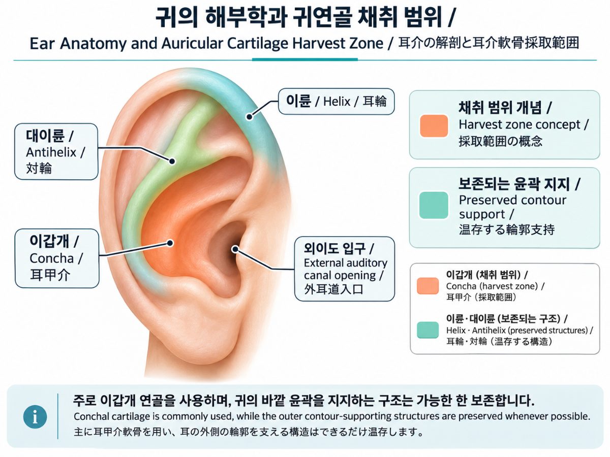
Which part of the ear is used in rhinoplasty?
The ear (auricle) is a thin layer of skin over a single, continuous piece of cartilage whose folds give the ear its form. In rhinoplasty, the portion most often used is the conchal cartilage in the central bowl. Because it is naturally curved, it suits purposes that call for curvature and some flexibility, such as refining the nasal tip, supporting the alar rim, or serving as small fillers.
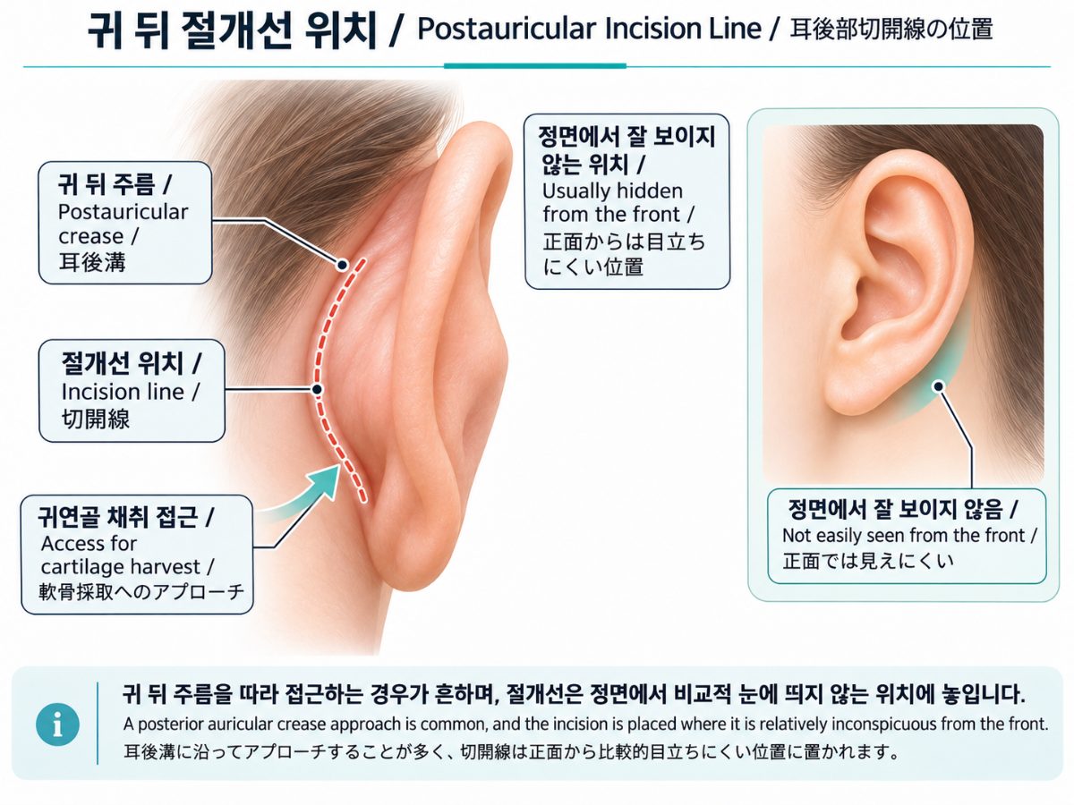
The outer rim (helix) and the ridge inside it (antihelix) act as the skeleton that determines the ear's shape, so they are preserved whenever possible. The extent of harvest is limited to what is needed, and the plan is generally made so as not to disturb the structures around the entrance to the ear canal excessively. A common approach is an incision along the crease behind the ear, which places the scar where it is hard to see from the front. The approach can vary, however, depending on what is needed and on the patient's ear anatomy.
What changes can appear in the ear after harvest?
It helps to separate changes that are common from those that are uncommon.
| Category | Change | Typical course (varies by individual) |
|---|---|---|
| Common | Swelling, bruising, a feeling of pressure, soreness | Usually settles over about 1–2 weeks |
| Common | Reduced sensation or a muffled feeling in the ear and surrounding skin | Recovers over weeks to months; in some people it can last longer |
| Common | A pink or red scar line behind the ear | Often fades over several months |
| Uncommon | Hematoma (collection of blood), infection | May require treatment if detected early |
| Uncommon | Hypertrophic scar or keloid | Strongly influenced by individual tendency; history must be checked |
| Uncommon | Subtle change in ear contour, or change in shape around the ear canal entrance | More likely when the harvested area is larger |
A change in hearing itself is generally not common, but excessive manipulation around the ear canal entrance can alter its shape, which is why the extent of harvest is limited at the planning stage. This is general information; the actual course differs from person to person.
How is the decision to harvest ear cartilage made in clinic?
① What is checked at the exam
When ear cartilage is being considered, the following are assessed together:
- Ear size, depth of the concha, cartilage thickness, and any asymmetry between the two ears
- History of ear surgery, injury, or piercing, and any skin inflammation or infection around the ear
- Tendency toward hypertrophic scarring or keloid (including how earlier surgical scars have healed)
- Daily pressure on the ear, such as glasses, earphones, and usual sleeping position
- The type and amount of graft required on the nasal side
② Criteria for choosing or avoiding a material
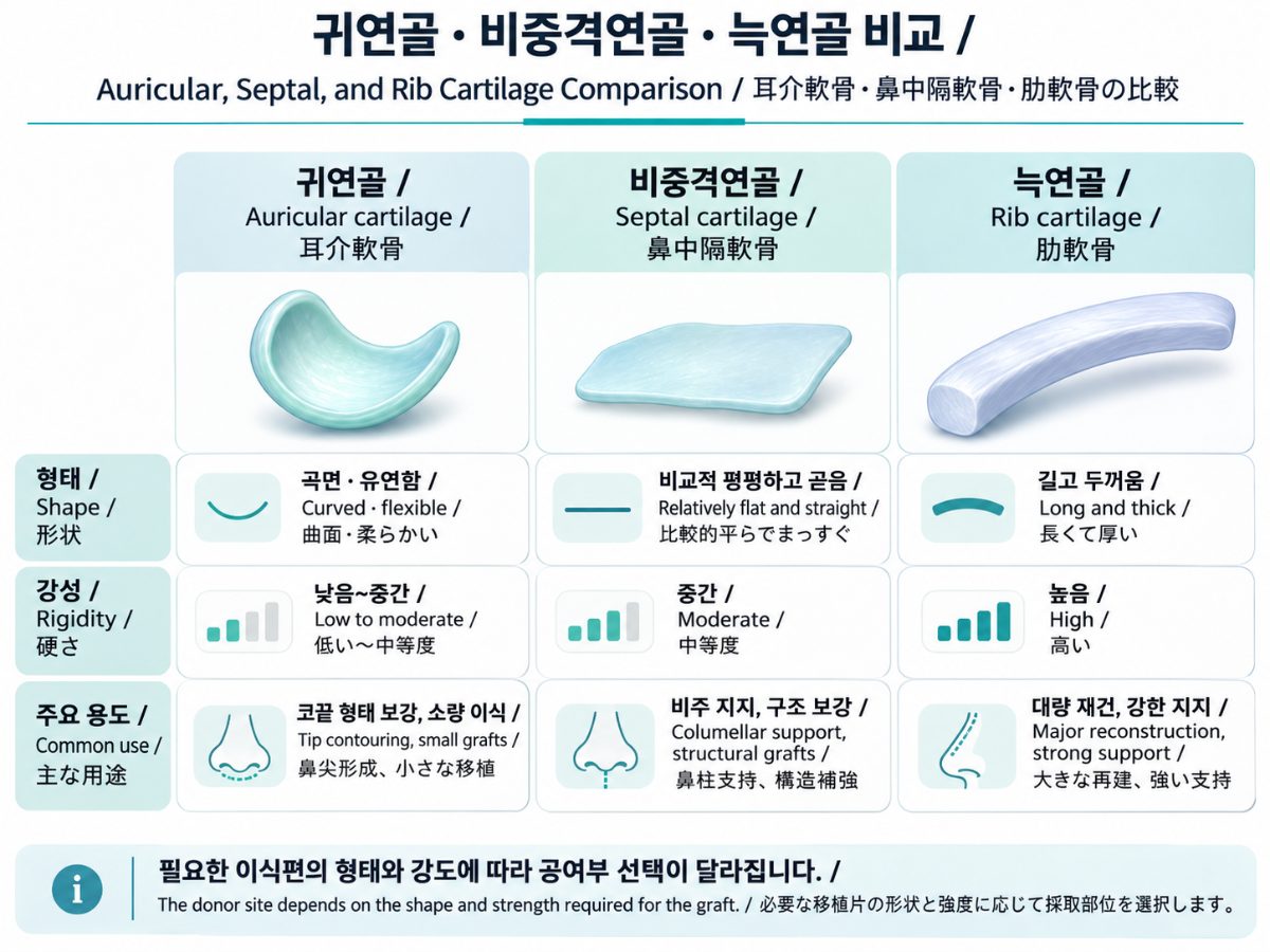
| Situation | Ear cartilage | Septal cartilage | Rib cartilage |
|---|---|---|---|
| Small amount needed, curvature and flexibility wanted (e.g., tip refinement) | May be considered | Possible if enough is available | Usually more than needed |
| Long, straight support needed (dorsal or columellar support) | Rigidity and length may be insufficient | Possible, but quantity is limited | Considered |
| Revision where septum is already used or insufficient | Considered as a supplementary donor site | Limited | Considered when a large amount is needed |
| Strong keloid tendency, or skin or infection problem of the ear | Deferred, or another donor site is considered | Not applicable | Not applicable |
Ear cartilage is curved, which makes it difficult to build long, straight structures from it, and the amount obtainable from one ear is limited. When a large supporting structure is needed, other donor sites are considered together rather than trying to solve the problem with ear cartilage alone. Conversely, when a small curved graft is enough, the judgment leans toward not choosing a larger donor site than necessary.
What does the decision process look like in practice?
The following is a hypothetical scenario to illustrate reasoning, not a patient's experience. Suppose a revision case shows weak tip support, and the septal cartilage has already been used with little left. The first step is to distinguish whether the graft needed is a small curved piece to refine the tip, or a supporting structure that needs length and rigidity. In the former case, after checking the ear anatomy and scarring tendency, conchal cartilage becomes a candidate; in the latter, ear cartilage alone may lack sufficient rigidity, and rib cartilage or another donor site is considered along with it. In this way, harvest is decided by weighing what is needed against how much burden that donor site carries.
Summary
- Ear cartilage is mainly taken from the concha, with the structures that support the ear's outline planned to be preserved.
- Swelling, bruising, and reduced sensation are relatively common; hematoma, infection, scarring, and contour change are uncommon but possible.
- It suits small, curved, flexible grafts, but has limits for long, straight supporting structures.
- Whether to harvest, and from where, depends on ear anatomy, scarring tendency, and the type and amount of graft needed, so individual assessment at an in-person consultation is necessary.
Frequently Asked Questions (FAQ)
Q1. Will the ear droop or change shape after harvest?
A. The helix and antihelix that support the ear's outline are planned to be preserved, so a noticeable change in appearance is not common. However, if the harvested area is large or the individual's cartilage is thin, a subtle change in contour is possible (varies by individual).
Q2. Is the scar behind the ear noticeable?
A. An incision along the crease behind the ear generally lies where it is hard to see from the front. Scar healing depends on constitution, though, so if you have a tendency toward hypertrophic scars or keloids, it is important to tell your surgeon beforehand.
Q3. Is a CT scan needed before harvesting ear cartilage?
A. The ear is generally assessed by inspection and palpation, and imaging is not always required. If the structure is uncertain because of past ear surgery or injury, additional evaluation may be considered.
Q4. Can cartilage be taken from both ears?
A. It can be considered depending on the amount needed, but the burden on the donor sites grows as more is taken. When a large structure is required, other donor sites are usually considered as well.
Related Columns
- Ear cartilage vs. septal cartilage: how do they differ in rhinoplasty?
- When Is Autologous Rib Cartilage Needed in Revision Rhinoplasty?
- Rib Cartilage in Rhinoplasty — Autologous vs. Donor Cartilage
- Why Does a Nasal Implant Show Through Thin Nose Skin?
Sources
- Korean Society of Plastic and Reconstructive Surgeons, Textbook of Plastic Surgery (chapters on rhinoplasty and cartilage grafting)
- Neligan PC (ed.), Plastic Surgery — rhinoplasty volume (Elsevier)
- Gunter JP et al. (eds.), Dallas Rhinoplasty: Nasal Surgery by the Masters (CRC Press)
- Daniel RK, Mastering Rhinoplasty (Springer)
Author · Reviewer · Dates
- Author: Dr. Jihun Kim, Board-Certified Plastic Surgeon (Chief Surgeon, VVLY Plastic Surgery)
- Published: 2026-11-04 · Last updated: 2026-11-04
※ This column provides general medical information and does not replace diagnosis or treatment for any individual patient. Indications, recovery, and possible complications vary between individuals, and an accurate assessment requires an in-person consultation with a specialist.